Author: admin

  • New Research Shows Air Pollution Could Play a Role in Development of Cardiometabolic Diseases, Diabetes

    New Research Shows Air Pollution Could Play a Role in Development of Cardiometabolic Diseases, Diabetes

    First-of-its-kind study, based on a mouse model, finds living in a polluted environment could be comparable to eating a high-fat diet, leading to a pre-diabetic state

    Air pollution is the world’s leading environmental risk factor, and causes more than nine million deaths per year. New research published in the Journal of Clinical Investigation shows air pollution may play a role in the development of cardiometabolic diseases, such as diabetes. Importantly, the effects were reversible with cessation of exposure.

    Researchers found that air pollution was a “risk factor for a risk factor” that contributed to the common soil of other fatal problems like heart attack and stroke. Similar to how an unhealthy diet and lack of exercise can lead to disease, exposure to air pollution could be added to this risk factor list as well.

    “In this study, we created an environment that mimicked a polluted day in New Delhi or Beijing,” said Sanjay Rajagopalan, MD, first author on the study, Chief of Cardiovascular Medicine at University Hospitals Harrington Heart and Vascular Institute, and Director of the Case Western Reserve University School of Medicine Cardiovascular Research Institute. “We concentrated fine particles of air pollution, called PM2.5 (particulate matter component < 2.5 microns). Concentrated particles like this develop from human impact on the environment, such as automobile exhaust, power generation and other fossil fuels.”

    These particles have been strongly connected to risk factors for disease. For example, cardiovascular effects of air pollution can lead to heart attack and stroke. The research team has shown exposure to air pollution can increase the likelihood of the same risk factors that lead to heart disease, such as insulin resistance and type 2 diabetes.

    In the mouse model study, three groups were observed: a control group receiving clean filtered air, a group exposed to polluted air for 24 weeks, and a group fed a high-fat diet. Interestingly, the researchers found that being exposed to air pollution was comparable to eating a high-fat diet. Both the air pollution and high-fat diet groups showed insulin resistance and abnormal metabolism – just like one would see in a pre-diabetic state.

    These changes were associated with changes in the epigenome, a layer of control that can masterfully turn on and turn off thousands of genes, representing a critical buffer in response to environmental factors. This study is the first-of-its-kind to compare genome-wide epigenetic changes in response to air pollution, compare and contrast these changes with that of eating an unhealthy diet, and examine the impact of air pollution cessation on these changes.

    “The good news is that these effects were reversible, at least in our experiments” added Dr. Rajagopalan. “Once the air pollution was removed from the environment, the mice appeared healthier and the pre-diabetic state seemed to reverse.”

    Dr. Rajagopalan explains that if you live in a densely polluted environment, taking actions such as wearing an N95 mask, using portable indoor air cleaners, utilizing air conditioning, closing car windows while commuting, and changing car air filters frequently could all be helpful in staying healthy and limiting air pollution exposure.

    Next steps in this research involve meeting with a panel of experts, as well as the National Institutes of Health, to discuss conducting clinical trials that compare heart health and the level of air pollution in the environment. For example, if someone has a heart attack, should they be wearing an N95 mask or using a portable air filter at home during recovery?

    Dr. Rajagopalan and his team believe that it is important to address the environment as a population health risk factor and continue to diligently research these issues. The authors also note that these findings should encourage policymakers to enact measures aimed at reducing air pollution.

    Shyam Biswal, PhD, Professor in the Department of Environmental Health and Engineering at Johns Hopkins University School of Public Health, is the joint senior author on the study. Drs. Rajagopalan and Biswal are co-PIs on the NIH grant that supported this work. 

    Rajagopalan, S., Biswal, S., et al. “Metabolic effects of air pollution exposure and reversibility.” Journal of Clinical Investigation. DOI: 10.1172/JCI137315.

    This work was supported by the National Institute of Environmental Health Sciences TaRGET II Consortium grant U01ES026721, as well as grants R01ES015146 and R01ES019616.

  • Babies Born to Depressed Moms Show Weakened Brain Connectivity

    Babies Born to Depressed Moms Show Weakened Brain Connectivity

    About one in five women experience some form of depression during pregnancy, with poorly understood effects on the fetus. Prenatal depression is linked to behavioural and developmental issues in children as well as an increased risk for depression as young adults. But how prenatal depression leads to these changes remains unclear.

    UCalgary researcher Dr. Catherine Lebel, PhD, is helping understand what may be happening in the developing brains of these children. The research team has shown that young children whose mothers experienced more numerous symptoms of depression in pregnancy have altered connectivity in brain pathways involved in emotion. These structural changes can be related to increased hyperactivity and aggression in boys.

    The research is based on diffusion magnetic resonance imaging, an imaging technique that probes the strength of structural connections between brain regions. The findings are published in The Journal of Neuroscience.

    Dr. Catherine Lebel, PhD

    Catherine Lebel, senior author and investigator.

    Riley Brandt, University of Calgary

    “The results help us understand how depression can have multigenerational impacts, and speaks to the importance of helping mothers who may be experiencing depression during pregnancy,” says Lebel, an associate professor at the Cumming School of Medicine, and researcher in the Alberta Children’s Hospital Research Institute. She holds the Canada Research Chair in Paediatric Neuroimaging.

    Lebel and her team studied 54 Calgary mothers and their children. They were enrolled from the ongoing, prospective study called the Alberta Pregnancy Outcomes and Nutrition study. Mothers answered a survey about their depression symptoms at several points during their pregnancy.

    Their children were followed after birth and undertook an MRI scan at the Alberta Children’s Hospital at around age four. As well, the children’s behaviour was assessed within six months of their MRI scan.

    The team found a significant reduction in structural brain connectivity between the amygdala, a structure essential for emotional processing, and the frontal cortex. Altered connectivity between the amygdala and frontal cortex is associated with disruptive behaviours and vulnerability to depression.

    The first author on the study, Dr. Rebecca Hay, MD, stresses the importance of recognition of depression and intervention in prenatal health. “These results suggest complex associations between the prenatal environment and children’s brain development, and may help us to understand why children of depressed mothers are more vulnerable to depression themselves,” says Hay, a resident physician in paediatrics and recent Cumming School of Medicine graduate.

    The main clinical takeaway from this is to emphasize the importance of recognizing, treating prenatal depression and supporting mothers, both for better maternal outcomes and to help future child development.

    Rebecca Hay

    Rebecca Hay, the study’s first author.

    Courtesy Rebecca Hay

    Current study looks at stress during pandemic

    Lebel and her research team are currently trying to understand how stress and mental health are affecting pregnant women during the COVID-19 pandemic. She is examining how factors such as social supports might mitigate stress, and how this may influence pregnancy and birth outcomes.

    If you are interested, you can get involved here in the Pregnancy During the COVID-19 Pandemic study at the University of Calgary. So far, approximately 7,500 women from across Canada are enrolled and supplying information through questionnaires.

    “It is critical to appropriately recognize and treat prenatal maternal mental health problems, both for the mothers and to improve child outcomes,” says Lebel. “Now more than ever, with increased stress, anxiety and depression during the COVID-19 pandemic, we should do more to support mothers to positively impact the health of their children.”

    Lebel is an associate professor in the Department of Radiology at the Cumming School of Medicine, adjunct associate professor in the Werklund School of Education and a member of The Mathison Centre for Mental Health Research & Education, Owerko Centre at ACHRI, Hotchkiss Brain Institute and Alberta Children’s Hospital Research Institute.

    The study was funded by the Canadian Institute of Health Research, Alberta Innovates – Health Solutions, the Alberta Children’s Hospital Foundation, the National Institute of Environmental Health Sciences, the Mach-Gaensslen Foundation, and an Eyes High University of Calgary Postdoctoral Scholar.

    Led by the Hotchkiss Brain Institute, Brain and Mental Health is one of six research strategies guiding the University of Calgary toward its Eyes High goals. The strategy provides a unifying direction for brain and mental health research at the university.

  • A New Tool for Modeling the Human Gut Microbiome

    A New Tool for Modeling the Human Gut Microbiome

    Several thousand strains of bacteria live in the human gut. Some of these are associated with disease, while others have beneficial effects on human health. Figuring out the precise role of each of these bacteria can be difficult, because many of them can’t be grown in lab studies using human tissue.

    This difficulty is especially pronounced for species that cannot live in oxygen-rich environments. However, MIT biological and mechanical engineers have now designed a specialized device in which they can grow those oxygen-intolerant bacteria in tissue that replicates the lining of the colon, allowing them to survive for up to four days.

    “We thought it was really important to contribute a tool to the community that could be used for this extreme case,” says Linda Griffith, the School of Engineering Professor of Teaching Innovation in MIT’s Department of Biological Engineering. “We showed that you can grow these very fastidious organisms, and we were able to study the effects they have on the human colon.”

    Using this system, the researchers showed that they could grow a strain of bacteria called Faecalibacterium prausnitzii, which lives in the human gut and protects against inflammation. They also showed that these bacteria, which are often diminished in patients with Crohn’s disease, appear to exert many of their protective effects through the release of a fatty acid called butyrate.

    Griffith and David Trumper, an MIT professor of mechanical engineering, are the senior authors of the study, which appears today in the journal Med. MIT postdocs Jianbo Zhang and Yu-Ja Huang are the lead authors of the paper.

    Oxygen sensitivity

    The human gut’s complex microbiome environment is difficult to model using animals such as mice, in part because mice eat a very different diet from humans, Griffith says.

    “We’ve learned a huge amount from mice and other animal models, but there are a lot of differences, especially when it comes to the gut microbiome,” she says.

    Most of the bacteria that live in the human gut are anaerobic, meaning that they do not require oxygen to survive. Some of these bacteria can tolerate low levels of oxygen, while others, such as F. prausnitzii, cannot survive oxygen exposure, which makes it difficult to study them in a laboratory. Some researchers have designed devices in which they can grow human colon cells along with bacteria that tolerate low levels of oxygen, but these don’t work well for F. prausnitzii and other highly oxygen-intolerant microbes.

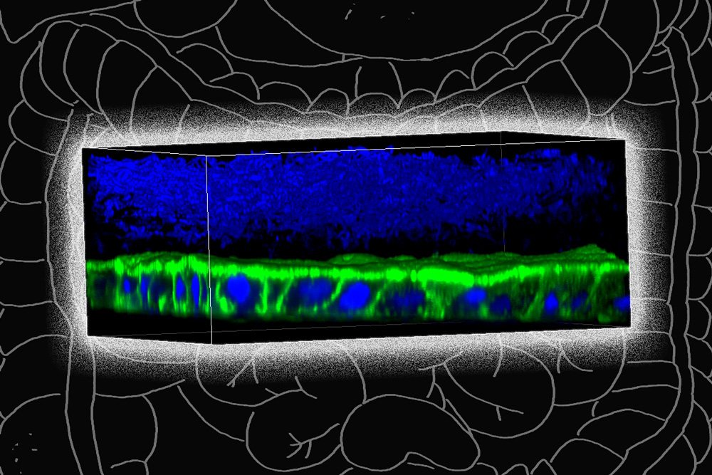
    To overcome this, the MIT team designed a device that allows them to precisely control oxygen levels in each part of the system. Their device contains a channel that is coated with cells from the human mucosal barrier of the colon. Below these cells, nutrients are pumped in to keep the cells alive. This bottom layer is oxygen-rich, but the concentration of oxygen decreases toward the top of the mucosal cell layer, similarly to what happens in the interior of the human colon.

    Just as they do in the human colon, the barrier cells in the channel secrete a dense layer of mucus. The MIT team showed that F. prausnitzii can form clouds of cells in the outer layer of this mucus and survive there for up to four days, in an environment that is kept oxygen-free by fluid flowing across it. This fluid also contains nutrients for the microbes.

    Using this system, the researchers were able to show that F. prausnitzii does influence cell pathways involved in inflammation. They observed that the bacteria produce a short-chain fatty acid called butyrate, which has previously been shown to reduce inflammation. After butyrate levels went up, the mucosal cells showed a reduction in the activity of a pathway called NF kappa B. This reduction calms inflammation.

    “Overall, this pathway has been reduced, which is really similar to what people have seen in humans,” Zhang says. “It seems that the bacteria are desensitizing the mammalian cells to not overreact to the dangers in the outside environment, so the inflammation status is being calmed down by the bacteria.”

    Patients with Crohn’s disease often have reduced levels of F. prausnitzii, and the lack of those bacteria is hypothesized to contribute to the overactive inflammation seen in those patients.

    When the researchers added butyrate to the system, without bacteria, it did not generate all of the effects that they saw when the bacteria were present. This suggests that some of the bacteria’s effects may be exerted through other mechanisms, which the researchers hope to further investigate.

    Microbes and disease

    The researchers also plan to use their system to study what happens when they add other species of bacteria that are believed to play a role in Crohn’s disease, to try to further explore the effects of each species.

    They are also planning a study, working with Alessio Fasano, the division chief of pediatric gastroenterology and nutrition at Massachusetts General Hospital, to grow mucosal tissue from patients with celiac disease and other gastrointestinal disorders. This tissue could then be used to study microbe-induced inflammation in cells with different genetic backgrounds.

    “We are hoping to get new data that will show how the microbes and the inflammation work with the genetic background of the host, to see if there could be people who have a genetic susceptibility to having microbes interfere with the mucosal barrier a little more than other people,” Griffith says.

    She also hopes to use the device to study other types of mucosal barriers, including those of the female reproductive tract, such as the cervix and the endometrium.

    The research was funded by the U.S. National Institutes of Health, the Boehringer Ingelheim SHINE Program, and the National Institute of Environmental Health Sciences.

    Press Mentions

    Nature

    Prof. Linda Griffith and her colleagues have “developed a model of the human gut to study how the organ’s microbes interact with immune cells and regulate inflammation,” reports Gemma Conroy for Nature. Griffith and her team “have also created models for endometriosis and pancreatic cancer,” writes Conroy. 

    Related Links

  • Major Climate Initiative in the Northeastern U.S. Benefits Children’s Health

    Major Climate Initiative in the Northeastern U.S. Benefits Children’s Health

    A new study by researchers from the Columbia Center for Children’s Environmental Health (CCCEH) at Columbia University Mailman School of Public Health reports that the Regional Greenhouse Gas Initiative (RGGI) has been successful in reducing fine particulate matter (PM2.5) emissions and substantially improving children’s health, both major co-benefits of this climate policy. Among the benefits between 2009-2014 were an estimated 537 avoided cases of childhood asthma, 112 preterm births, 98 cases of autism spectrum disorder (ASD), and 56 cases of term low birthweight. The associated economic savings were estimated at $191 to $350 million over the years. These findings are published today in Environmental Health Perspectives.

    Initiated in 2009, RGGI is the country’s first regional cap- and- trade program designed to limit carbon dioxide (CO2) emissions from the power sector. Under the agreement, nine participating northeastern states—including New York— were required to limit emissions from fossil fuel power plants with a capacity of 25 megawatts or greater. Regulated power plants must obtain annual CO2 allowances which are auctioned quarterly and may be traded between plants or offset. Although RGGI is focused on reducing GHG emissions, it has also lowered emissions of other pollutants, including PM2.5., nitrogen oxides (NOx) and sulfur dioxide (SO2).

    The researchers estimated the health benefits to children and associated economic savings using the Environmental Benefits Mapping and Analysis Program (BenMAP) tool, a computer program supported by the U.S. Environmental Protection Agency. For the first time, the researchers incorporated additional health outcomes, not previously included in BenMAP, that are associated with prenatal and childhood exposure to PM2.5.

    “As impressive as they are, these estimated benefits for children do not take into account their potential life-long consequences, so they are likely underestimates of the true benefits of this policy,” says lead author Frederica Perera, PhD, DrPH, professor of environmental health sciences at Columbia Mailman School and director of translational research at CCCEH. “These results should spur more such initiatives to address climate change and improve the health of our children.”

    Additional authors include David Cooley at Abt Associates, Alique Berberian at the Columbia Center for Children’s Environmental Health, David Mills at Peak to Peak Economics, and Patrick Kinney at Boston University’s School of Public Health.

    This research was supported by the Environmental Protection Agency (RD83615401) and National Institute of Environmental Health Sciences (P50ES09600), the John Merck Fund, the John and Wendy Neu Family Foundation, and an anonymous donor.

  • Higher BPA Levels Linked to More Asthma Symptoms in Children

    Children in low-income neighborhoods in Baltimore tended to have more asthma symptoms when levels of the synthetic chemical BPA (Bisphenol A) in their urine were elevated, according to a study from researchers at the Johns Hopkins Bloomberg School of Public Health and School of Medicine.

  • Study: Novel PFAS Comprise 24% of Those Measured in Blood of Wilmington, N.C. Residents

    Study: Novel PFAS Comprise 24% of Those Measured in Blood of Wilmington, N.C. Residents

    In a new paper detailing findings from North Carolina State University’s GenX Exposure Study, researchers detected novel per- and polyfluoroalkyl substances (PFAS) called “fluoroethers” in blood from residents of Wilmington, North Carolina. The fluoroethers – Nafion byproduct 2, PFO4DA and PFO5DoA – represented 24% of the total PFAS detected in the blood of Wilmington residents and appear to leave the body faster than legacy PFAS. These are the first measurements of these chemicals in humans.

    The GenX Exposure Study began in 2017 after NC State researchers found a chemical called GenX in Wilmington residents’ drinking water. An upstream chemical facility had been releasing several PFAS (including GenX) into the Cape Fear River, the city’s primary drinking water source, since 1980. The study aimed to answer community questions about GenX and other PFAS, including whether the chemicals were detectable in residents’ blood.

    NC State and East Carolina University researchers collected blood samples from 344 Wilmington residents (289 adults and 55 children) across two sampling efforts in November 2017 and May 2018. Additionally, 44 of the November participants had a second blood sample collected in May 2018, six months after their first one. All blood samples were collected after the chemical facility stopped releasing GenX into the Cape Fear River.

    The first sample determined which PFAS were present and the second sample was used to see how the levels changed in six months. The study looked for 10 PFAS unique to the chemical facility (called “fluoroethers”), and 10 legacy PFAS (such as PFOA and PFOS), in the samples.

    Ten PFAS were found in most of the blood samples. Three of these were the fluoroethers Nafion byproduct 2, PFO4DA and PFO5DoA, all of which were detected in over 85% of samples. Two other fluoroethers, PFO3OA and NVHOS, were infrequently detected. Nearly all samples had at least one fluoroether present, but GenX was not found in any samples.

    In total, 24% of the measured PFAS in blood samples came from these novel fluoroethers. In participants with repeated samples, the median decrease in fluoroether levels ranged from 28% for PFO5DoA to 65% for PFO4DA in six months once exposure to contaminants in drinking water ceased.

    Four legacy PFAS (PFHxS, PFOA, PFOS, PFNA) were detected in most (97% or greater) participants. The levels of legacy PFAS in the participants’ blood were higher than U.S. national levels for the 2015-2016 National Health and Nutrition Examination Survey and did not change in the repeated samples.

    “Accurately measuring the levels of PFAS in blood is the first step toward understanding what effects these novel chemicals may have on human health,” says Jane Hoppin, professor of biological sciences, deputy director of NC State’s Center for Human Health and the Environment (CHHE), and corresponding author of the paper describing the work.

    The study appears in Environmental Health Perspectives and is supported by the National Institute of Environmental Health Sciences (1R21ES029353), NC State’s Center for Human Health and the Environment (P30ES025128), and the NC Policy Collaboratory. Partners include East Carolina University, the Environmental Protection Agency, New Hanover County Health Department and Cape Fear River Watch.

    -peake-

    Note to editors: An abstract follows.

    “Measurement of novel, drinking water-associated PFAS in blood from adults and children in Wilmington, North Carolina”

    DOI: 10.1289/EHP6837

    Authors: Nadine Kotlarz, Katlyn May, Robert Smart, Detlef Knappe, Jane Hoppin; North Carolina State University; James McCord, Mark Strynar, Andrew Lindstrom, U.S. Environmental Protection Agency; David Collier, Suzanne Lea, Jamie DeWitt, East Carolina University; Adrien A. Wilkie, Jessica Y. Islam, UNC Chapel Hill; Katelyn Matney, Phillip Tarte, New Hanover County Health Department; M.E. Polera, Kemp Burdette, Cape Fear River Watch
    Published: Online in Environmental Health Perspectives

    Abstract:
    BACKGROUND: From 1980 to 2017, a fluorochemical manufacturing facility discharged wastewater containing poorly understood per- and polyfluoroalkyl substances (PFAS) to the Cape Fear River, the primary drinking water source for Wilmington, North Carolina (NC), residents. Those PFAS included several fluoroethers including HFPO-DA or “GenX”. Little is known about the bioaccumulation potential of these fluoroethers.
    OBJECTIVE: To determine levels of fluoroethers and legacy PFAS in serum from Wilmington, NC, residents.
    METHODS: In November 2017 and May 2018, we enrolled 344 Wilmington, NC, residents aged 6 years and older into the GenX Exposure Study, and collected blood samples. Repeated blood samples were collected from 44 participants six months after enrollment. We analyzed serum for 10 fluoroethers and 10 legacy PFAS using liquid chromatography-high resolution mass spectrometry.
    RESULTS: Participants’ ages ranged from 6 to 86 years; they lived in the lower Cape Fear region for 20 years on average (standard deviation: 16 years). Six fluoroethers were detected in serum; Nafion byproduct 2, PFO4DA, and PFO5DoA were detected in >85% of participants. PFO3OA and NVHOS were infrequently detected. Hydro-EVE was present in a subset of samples but we could not quantify it. GenX was not detected above our analytical method reporting limit (2 ng/mL). In participants with repeated samples, the median decrease in fluoroether levels ranged from 28% for PFO5DoA to 65% for PFO4DA in six months due to wastewater discharge control. Four legacy PFAS (PFHxS, PFOA, PFOS, PFNA) were detected in most (≥ 97%) participants; these levels were higher than U.S. national levels for 2015-2016 National Health and Nutrition Examination Survey. The sum concentration of fluoroethers contributed 24% to participants’ total serum PFAS (median: 25.3 ng/mL).
    CONCLUSION: Poorly understood fluoroethers released into the Cape Fear River by a fluorochemical manufacturing facility were detected in blood from Wilmington, NC, residents. Health implications of exposure to these novel PFAS have not been well characterized.

  • Jet Aircraft Exhaust Linked to Preterm Births

    Jet Aircraft Exhaust Linked to Preterm Births

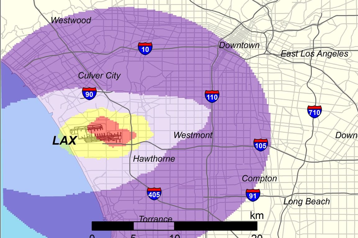
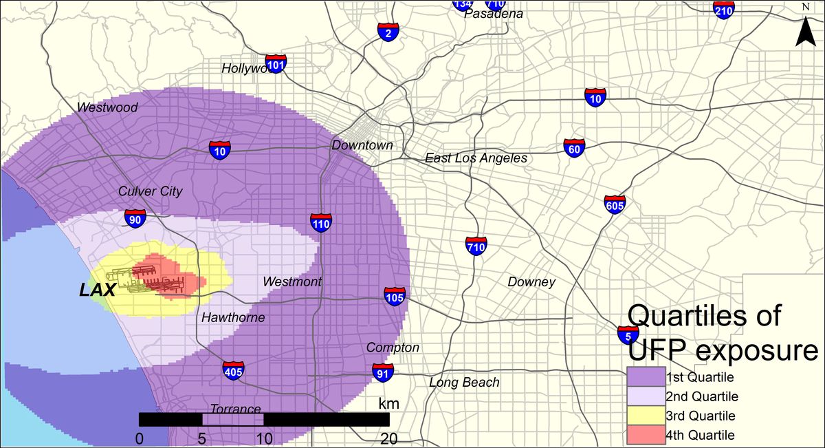
    Map showing levels of pollution from jet exhaust

    UCLA Fielding School of Public Health

    UCLA researchers found that mothers who lived within the red area adjacent to LAX were 14% more likely to have a pregnancy that ended in a preterm birth. Those in the yellow area were 8% percent more likely than those in areas farther from the airport.

    A study from the UCLA Fielding School of Public Health has found that pregnant women exposed to high levels of ultrafine particles from jet airplane exhaust are 14% more likely to have a preterm birth than those exposed to lower levels.

    The researchers examined exposure among women living near Los Angeles International Airport, in an area that includes neighborhoods in Los Angeles, El Segundo, Hawthorne, Inglewood and several other communities inland from the airport.

    “The data suggest that airplane pollution contributes to preterm births above and beyond the main source of air pollution in this area, which is traffic,” said Dr. Beate Ritz, a professor in the departments of epidemiology and environmental health sciences at the Fielding School.

    Preterm birth is associated with complications such as immature lungs, difficulty regulating body temperature, poor feeding and slow weight gain.

    The research team, co-led by Ritz and Scott Fruin of the Keck School of Medicine at the University of Southern California, examined records for all births — a total of 174,186 —between 2008 and 2016 to mothers living within 9 miles (15 km) of LAX. They divided the overall area into four sections based on the amount of ultrafine particle, or UFP, pollution from jet exhaust, with the section nearest the airport experiencing the highest exposure.

    After adjusting for traffic-related air pollution and other variables that may affect the risk of preterm birth, including airport-related noise and the mother’s age, education level and race, they found that expectant mothers in the quarter with the highest average ultrafine particle exposure had 14% higher odds of a preterm birth than mothers in the quarter with the lowest exposure.

    “Nearly 2 million people live within a 10–mile radius of LAX, many of whom are exposed to elevated levels of aircraft-origin UFPs,” said Sam Wing, a scholar at the Fielding School who also worked on the study.

    The research is summarized in an article published July 22 in the journal Environmental Health Perspectives and is based on a lengthy study from the spring 2020 edition of the journal. Other authors include Timothy Larson and Sarunporn Boonyarattaphan of the University of Washington and Neelakshi Hudda of Tufts University.

    This research was supported by grants from the National Institute of Environmental Health Sciences, part of the National Institutes of Health.

  • Living Near Natural Gas Flaring Poses Health Risks for Pregnant Women and Babies

    Living Near Natural Gas Flaring Poses Health Risks for Pregnant Women and Babies

    Natural gas flares can burn for weeks at a time. (Photo/iStock)

    Natural gas flares can burn for weeks at a time. (Photo/iStock)

    Health

    USC researchers found that exposure to flaring was associated with 50% higher odds of preterm birth.

    July 15, 2020 Leigh Hopper

    Researchers from USC and UCLA have found that women living near natural gas and oil wells that use flaring to burn off excess gas face a 50% greater risk of premature birth than women with no exposure.

    “Our study finds that living near flaring is harmful to pregnant women and babies,” said Jill Johnston, an environmental health scientist at the Keck School of Medicine of USC. “We have seen a sharp increase in flaring in Texas’ Eagle Ford Shale, and this is the first study to explore the potential health impacts.”

    Flares, which can burn for weeks at a time, release harmful chemicals such as benzene as well as fine particle pollution, carbon monoxide, nitrogen oxides, heavy metals and black carbon. Several of these combustion-related pollutants are linked to a higher risk of preterm birth and reduced birth weight in other contexts.

    The research appears today in the journal Environmental Health Perspectives.

    Exposure to natural gas flaring associated with preterm birth

    The study examined 23,487 live births to women living within the Eagle Ford region between 2012 to 2015. The Eagle Ford Shale geological formation, measuring 50 miles wide and 400 miles long, is one of the most productive oil and gas regions in the country due to hydraulic fracturing, or “fracking.” In a previous study, the research team estimated the area was subject to more than 43,000 flaring events between 2012 and 2016.

    The preterm birth rate was 14% among pregnant women exposed to a high number of flares, researchers found. Babies born prematurely — before the 37th completed week of pregnancy — may suffer complications such as immature lungs, difficulty regulating body temperature, poor feeding and slow weight gain.

    Our study finds that living near flaring is harmful to pregnant women and babies.

    Jill Johnston

    The researchers used satellite observations to measure flaring activity because systemic reporting is lacking. They adjusted for other known risk factors for preterm birth in their analysis — including age, smoking, insurance status and access to prenatal care — and concluded that exposure to a high amount of flaring was associated with 50% higher odds of preterm birth compared with no exposure. A high amount of flaring was defined as 10 or more nightly flare events within 3 miles of the pregnant woman’s home.

    “Women who identified as Latina or Hispanic in our study were exposed to more flaring and more likely to see an increased risk of preterm birth, raising environmental justice concerns about the oil and gas boom in south Texas,” said Lara Cushing, an environmental health scientist with the UCLA Fielding School of Public Health who co-led the study with Johnston. “Our study adds to the evidence that oil and gas development is negatively impacting birth outcomes and suggests stricter regulation of the industry is needed.”

    Exposure to wells, race also contribute to higher odds of preterm birth

    Women who lived within 3 miles of a high number of oil and gas wells also had higher odds of preterm birth than mothers who did not live near wells. Their babies were also born weighing 19.4 grams, or seven ounces, lighter on average. This suggests that, in addition to flaring, other exposures related to oil and gas wells may also be adversely impacting pregnancy, the researchers said.

    The majority (55%) of the women in the study population identified as Latina or Hispanic, and the odds of preterm birth among Hispanic women exposed to high levels of flaring was greater than the corresponding odds among non-Hispanic White women, who made up 37% of the study population. Nearly 60% of women in the study were on public health insurance (Medicaid) and 17% were foreign born.

    Amid an oil boom in recent years, the United States has been responsible for the highest number of flares of any country, flaring an estimated 14.1 billion square meters of natural gas in 2018. Eighty percent of flaring is occurring in Texas and in North Dakota shale plays, where much of the U.S. fracking occurs. That said, according to researchers, flaring largely remains underreported and unregulated.


    In addition to Johnston and Cushing, other authors of the study include Kate Vavra-Musser, Khang Chau and Meredith Franklin, all of USC.

    The study was supported by a grant from the U.S. National Institutes of Health/National Institute of Environmental Health Sciences.

  • Bed Bugs Modify Microbiome of Homes They Infest

    Bed Bugs Modify Microbiome of Homes They Infest

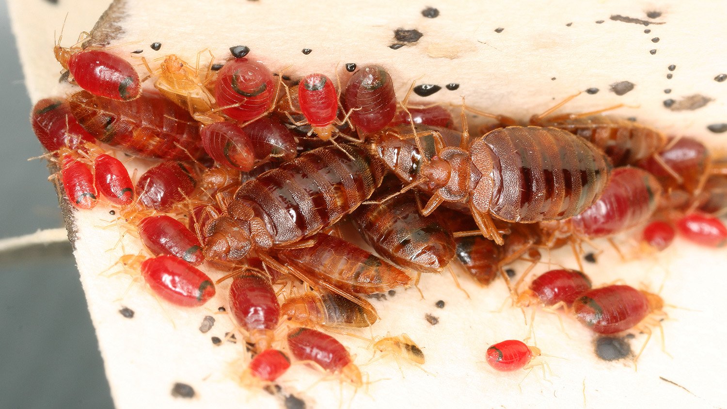
    Homes infested by bed bugs appear to have different bacterial communities – often referred to as microbiomes – than homes without bed bugs, according to a first-of-its-kind study from North Carolina State University. In addition, once bed bug infestations were eradicated, home microbiomes became more similar to those in homes that didn’t have bed bugs. The findings could be an important step in lifting the veil on the factors involved in indoor environmental quality and how to improve it.

    Microbes can affect indoor air quality. So NC State entomologists Coby Schal and Madhavi Kakumanu wanted to learn more about the microbiomes of bed bugs, whether bed bugs can shape the microbial community in homes they infest, and whether eliminating bed bugs changes the microbiome of homes that were once infested.

    The study, held in an apartment complex in Raleigh, compared the microbiomes of bed bugs with the microbiomes in the household dust of infested homes as well as the microbiomes in apartments that had no bed bugs. Nineteen infested homes were studied over the course of four months; seven were treated with heat to eliminate bed bugs after the initial sample was taken, while 12 infested homes were treated after one month. These homes were compared with 11 homes that had no bed bugs.

    The results showed similarities between the microbiomes of bed bugs and the dust-associated microbiomes of infested homes, mostly through the presence of Wolbachia, a symbiotic bacterium that comprises the majority of the bacterial abundance in bed bugs. Bed bug and infested home microbiomes differed significantly from the microbial communities of uninfested homes.

    “There is a link between the microbiome of bed bugs and the microbiome of household dust in bed bug infested homes,” said Schal, the Blanton J. Whitmire Distinguished Professor of Entomology at NC State and co-corresponding author of the paper. “No previous study has reported the impact of chronic pest infestations on indoor microbial diversity.”

    The study also showed that, after bed bugs were eliminated, infested home microbiomes gradually became more like those in homes without bed bugs.

    “The elimination of the bed bugs resulted in gradual shifts in the home microbial communities toward those of uninfested homes,” Kakumanu, an NC State research scholar in Schal’s lab and co-corresponding author of the study, said. “This paper is the first experimental demonstration that eliminating an indoor pest alters the indoor microbiome toward that of uninfested homes.”

    “Bed bug infestations are problematic in many homes in both developed and developing countries,” Schal said. “There is a critical need to investigate infestations from the perspective of indoor environmental quality, and this paper represents a first step toward this end.” 

    The study appears in Science of the Total Environment. Funding for the work came from NC State’s Blanton J. Whitmire endowment, as well as the U.S. Department of Housing and Urban Development Healthy Homes program (NCHHU0017-13, NCHHU0053-19), the Alfred P. Sloan Foundation (2013-5-35 MBE) and the National Science Foundation (DEB-1754190). Seed funds came from NC State’s Center for Human Health and the Environment (CHHE, P30ES025128), funded by the National Institute of Environmental Health Sciences.

    -kulikowski-

    Note to editors: An abstract of the paper follows.

    “Bed bugs shape the indoor microbial community composition of infested homes”
    Authors: Madhavi L. Kakumanu, Alexis M. Barbarin, Richard G. Santangelo and Coby Schal, North Carolina State University: Zachary C. DeVries, University of Kentucky

    Published: July 7, 2020 in Science of the Total Environment

    DOI: 10.1016/j.scitotenv.2020.140704

    Abstract: Indoor pests, and the allergens they produce, adversely affect human health. Surprisingly, however, their effects on indoor microbial communities have not been assessed. Bed bug (Cimex lectularius) infestations pose severe challenges in elderly and low-income housing. They void large amounts of liquid feces into the home environment, which might alter the indoor microbial community composition. In this study, using bed bug-infested and uninfested homes, we showed a strong impact of bed bug infestations on the indoor microbial diversity. Floor dust samples were collected from uninfested and bed bug-infested homes and their microbiomes were analyzed before and after heat interventions that eliminated bed bugs. The microbial communities of bed bug-infested homes were radically different from those of uninfested homes, and the bed bug endosymbiont Wolbachia was the major driver of this difference. After bed bugs were eliminated, the microbial community gradually shifted toward the community composition of uninfested homes, strongly implicating bed bugs in shaping the dust-associated environmental microbiome. Further studies are needed to understand the viability of these microbial communities and the potential risks that bed bug-associated microbes and their metabolites pose to human health.

  • Childhood Obesity Linked to Multiple Environmental Factors in First-of-its-kind Study

    Childhood Obesity Linked to Multiple Environmental Factors in First-of-its-kind Study

    Childhood obesity is a health threat that is becoming more and more common worldwide. It increases risk later on for a variety of life-threatening challenges, including type 2 diabetes, cancer, heart disease and even mental health problems.

    A new study led by scientists at USC and the Barcelona Institute for Global Health (ISGlobal) is the first to comprehensively profile environmental factors linked to childhood obesity. The research showed that a higher body mass index (BMI), an estimate of body fat, during childhood is associated with exposure to smoking — both in the womb and while growing up — as well as air pollution and certain characteristics seen in some urban areas. Differences in socioeconomic status did not explain these results.

    The findings were just published in the journal Environmental Health Perspectives.

    “People are not exposed to only one chemical during their lives,” said Leda Chatzi, MD, PhD, associate professor of preventive medicine at the Keck School of Medicine of USC and the senior author of the study. “They are exposed to multiple chemicals. With that in mind, we try to understand the totality of environmental exposures.”

    Dr. Chatzi and her colleagues are exploring the exposome, an endeavor meant to complement investigations into the genome. Just as many studies of the genome attempt to clarify how the content of genes affect health, studies such as this one attempt to clarify how health is affected by every environmental influence from conception onward. This new approach contrasts with a traditional method for probing population health, zeroing in on one or two environmental factors in a given investigation.

    All told, the paper examined 173 factors — 77 during pregnancy and 96 during childhood. These included air pollutants, families’ human-made surroundings and access to green space, tobacco smoke and chemical pollutants such as heavy metals and pesticides.

    The researchers studied a group of about 1,300 children aged 6 to 11 years from six European countries: France, Greece, Lithuania, Norway, Spain and the United Kingdom. Data about the women and their children have been gathered, starting at pregnancy, through a collaborative longitudinal research project known as the Human Early Life Exposome (HELIX) study.

    Smoking exposure, air pollution and lack of exercise

    Mothers’ smoking during pregnancy was the most prominent association with high BMI among children, and the only prenatal factor with a significant association. Additionally, high BMI was associated with exposure to secondhand smoke, as measured through levels of a certain chemical in childrens’ urine samples. Taken together, these findings suggest that kicking the habit — or never picking it up — is one way that parents can safeguard the long-term health of their offspring.

    “This is quite an important message,” Dr. Chatzi said. “Maternal smoking during pregnancy and exposures to secondhand smoke are quite prevalent worldwide.”

    Exposure to air pollution, both indoors and out, was another factor linked to higher BMI. Specific pollutants were nitrogen dioxide — a component of automobile exhaust and other gases released when fossil fuels burn — as well as particles in the atmosphere.

    Certain attributes of the areas where children live also showed a strong correlation with BMI. BMI was higher for children who live in densely populated areas. But BMI was lower for those who went to school in areas that are denser in facilities such as businesses, community services, educational institutions, restaurants and shopping — a proxy for a neighborhood’s walkability.

    “With more facilities, children can walk, ride their bikes, go play sports,” Dr. Chatzi said. “You can contrast this with what are described as food deserts, or areas with fewer facilities.”

    The researchers note that better understanding of the impact of environmental exposure could create opportunities to take action that reverses the trend of increasing childhood obesity, ultimately mitigating its long-term dangers.

    “These findings provide further evidence that modifying environmental exposures early in life can limit the risk of obesity and associated complications,” said first author Martine Vrijheid, research professor at ISGlobal and principal investigator of the HELIX project. “The implications for public health are important since these results may help to identify obesity-related exposures that could be targeted for prevention and intervention early in life.”

    About this study                                                 

    In addition to Dr. Chatzi and Vrijheid, the studies’ co-authors are Serena Fossati, Léa Maitre, Sandra Márquez, Maribel Casas, Montserrat de Castro,  David Donaire-Gonzalez, Oliver Robinson, Jordi Sunyer, Ibon Tamayo-Uria, Jose Urquiza, Antonia Valentin, Charline Warembourg, Mark J. Nieuwenhuijsen and Xavier Basagaña from the the ISGlobal Institute, Barcelona, Spain; Theano Roumeliotaki and Marina Vafeiadi from the Department of Social Medicine, Faculty of Medicine, University of Crete, Heraklion, Crete, Greece; Lydiane Agier, Solène Cadiou and Valerie Siroux from the Team of Environmental Epidemiology Applied to Reproduction and Respiratory Health, INSERM, CNRS, University Grenoble Alpes, Institute for Advanced Biosciences (IAB), U1209 Joint Research Center, Grenoble, France; Sandra Andrusaityte, Audrius Dedele and Regina Grazuleviciene from the Department of Environmental Sciences, Vytautas Magnus University, Kaunas, Lithuania; Line S. Haug, Helle Margrete Meltzer, Eleni Papadopouplou, Amrit K. Sakhi, Per E. Schwarze and Cathrine Thomsen from the Norwegian Institute of Public Health, Oslo, Norway; and Rosemary McEachan and  John Wright from the Bradford Institute for Health Research, Bradford Teaching Hospitals NHS Foundation Trust, Bradford, UK.

    This study was supported by the European Community Seventh Framework Program (Human Early Life Exposome Project, grant ID: 308333) and by the National Institute of Environmental Health Sciences (R21ES029681).

    — by Wayne Lewis