Author: admin

  • A New Tool for Modeling the Human Gut Microbiome

    A New Tool for Modeling the Human Gut Microbiome

    Several thousand strains of bacteria live in the human gut. Some of these are associated with disease, while others have beneficial effects on human health. Figuring out the precise role of each of these bacteria can be difficult, because many of them can’t be grown in lab studies using human tissue.

    This difficulty is especially pronounced for species that cannot live in oxygen-rich environments. However, MIT biological and mechanical engineers have now designed a specialized device in which they can grow those oxygen-intolerant bacteria in tissue that replicates the lining of the colon, allowing them to survive for up to four days.

    “We thought it was really important to contribute a tool to the community that could be used for this extreme case,” says Linda Griffith, the School of Engineering Professor of Teaching Innovation in MIT’s Department of Biological Engineering. “We showed that you can grow these very fastidious organisms, and we were able to study the effects they have on the human colon.”

    Using this system, the researchers showed that they could grow a strain of bacteria called Faecalibacterium prausnitzii, which lives in the human gut and protects against inflammation. They also showed that these bacteria, which are often diminished in patients with Crohn’s disease, appear to exert many of their protective effects through the release of a fatty acid called butyrate.

    Griffith and David Trumper, an MIT professor of mechanical engineering, are the senior authors of the study, which appears today in the journal Med. MIT postdocs Jianbo Zhang and Yu-Ja Huang are the lead authors of the paper.

    Oxygen sensitivity

    The human gut’s complex microbiome environment is difficult to model using animals such as mice, in part because mice eat a very different diet from humans, Griffith says.

    “We’ve learned a huge amount from mice and other animal models, but there are a lot of differences, especially when it comes to the gut microbiome,” she says.

    Most of the bacteria that live in the human gut are anaerobic, meaning that they do not require oxygen to survive. Some of these bacteria can tolerate low levels of oxygen, while others, such as F. prausnitzii, cannot survive oxygen exposure, which makes it difficult to study them in a laboratory. Some researchers have designed devices in which they can grow human colon cells along with bacteria that tolerate low levels of oxygen, but these don’t work well for F. prausnitzii and other highly oxygen-intolerant microbes.

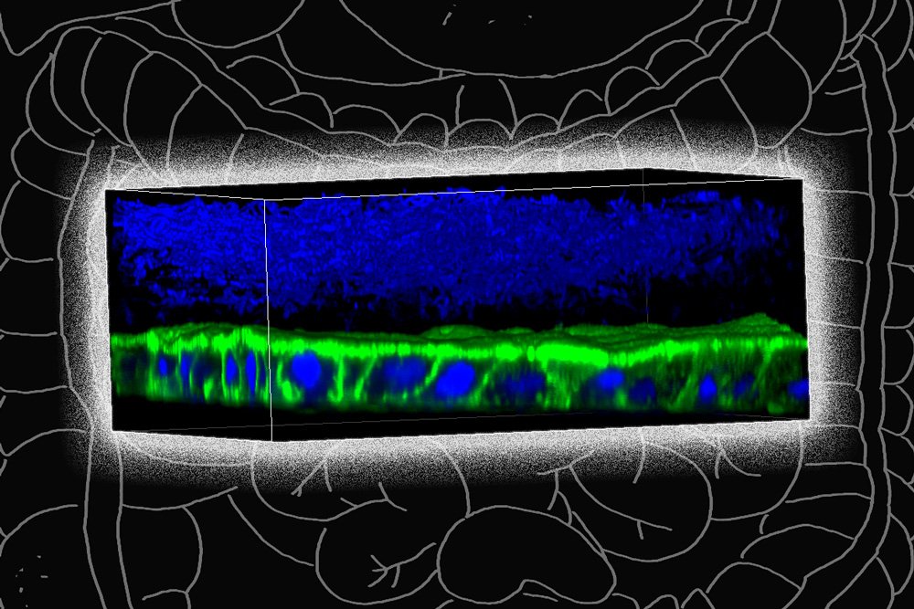
    To overcome this, the MIT team designed a device that allows them to precisely control oxygen levels in each part of the system. Their device contains a channel that is coated with cells from the human mucosal barrier of the colon. Below these cells, nutrients are pumped in to keep the cells alive. This bottom layer is oxygen-rich, but the concentration of oxygen decreases toward the top of the mucosal cell layer, similarly to what happens in the interior of the human colon.

    Just as they do in the human colon, the barrier cells in the channel secrete a dense layer of mucus. The MIT team showed that F. prausnitzii can form clouds of cells in the outer layer of this mucus and survive there for up to four days, in an environment that is kept oxygen-free by fluid flowing across it. This fluid also contains nutrients for the microbes.

    Using this system, the researchers were able to show that F. prausnitzii does influence cell pathways involved in inflammation. They observed that the bacteria produce a short-chain fatty acid called butyrate, which has previously been shown to reduce inflammation. After butyrate levels went up, the mucosal cells showed a reduction in the activity of a pathway called NF kappa B. This reduction calms inflammation.

    “Overall, this pathway has been reduced, which is really similar to what people have seen in humans,” Zhang says. “It seems that the bacteria are desensitizing the mammalian cells to not overreact to the dangers in the outside environment, so the inflammation status is being calmed down by the bacteria.”

    Patients with Crohn’s disease often have reduced levels of F. prausnitzii, and the lack of those bacteria is hypothesized to contribute to the overactive inflammation seen in those patients.

    When the researchers added butyrate to the system, without bacteria, it did not generate all of the effects that they saw when the bacteria were present. This suggests that some of the bacteria’s effects may be exerted through other mechanisms, which the researchers hope to further investigate.

    Microbes and disease

    The researchers also plan to use their system to study what happens when they add other species of bacteria that are believed to play a role in Crohn’s disease, to try to further explore the effects of each species.

    They are also planning a study, working with Alessio Fasano, the division chief of pediatric gastroenterology and nutrition at Massachusetts General Hospital, to grow mucosal tissue from patients with celiac disease and other gastrointestinal disorders. This tissue could then be used to study microbe-induced inflammation in cells with different genetic backgrounds.

    “We are hoping to get new data that will show how the microbes and the inflammation work with the genetic background of the host, to see if there could be people who have a genetic susceptibility to having microbes interfere with the mucosal barrier a little more than other people,” Griffith says.

    She also hopes to use the device to study other types of mucosal barriers, including those of the female reproductive tract, such as the cervix and the endometrium.

    The research was funded by the U.S. National Institutes of Health, the Boehringer Ingelheim SHINE Program, and the National Institute of Environmental Health Sciences.

    Press Mentions

    Nature

    Prof. Linda Griffith and her colleagues have “developed a model of the human gut to study how the organ’s microbes interact with immune cells and regulate inflammation,” reports Gemma Conroy for Nature. Griffith and her team “have also created models for endometriosis and pancreatic cancer,” writes Conroy. 

    Related Links

  • Major Climate Initiative in the Northeastern U.S. Benefits Children’s Health

    Major Climate Initiative in the Northeastern U.S. Benefits Children’s Health

    A new study by researchers from the Columbia Center for Children’s Environmental Health (CCCEH) at Columbia University Mailman School of Public Health reports that the Regional Greenhouse Gas Initiative (RGGI) has been successful in reducing fine particulate matter (PM2.5) emissions and substantially improving children’s health, both major co-benefits of this climate policy. Among the benefits between 2009-2014 were an estimated 537 avoided cases of childhood asthma, 112 preterm births, 98 cases of autism spectrum disorder (ASD), and 56 cases of term low birthweight. The associated economic savings were estimated at $191 to $350 million over the years. These findings are published today in Environmental Health Perspectives.

    Initiated in 2009, RGGI is the country’s first regional cap- and- trade program designed to limit carbon dioxide (CO2) emissions from the power sector. Under the agreement, nine participating northeastern states—including New York— were required to limit emissions from fossil fuel power plants with a capacity of 25 megawatts or greater. Regulated power plants must obtain annual CO2 allowances which are auctioned quarterly and may be traded between plants or offset. Although RGGI is focused on reducing GHG emissions, it has also lowered emissions of other pollutants, including PM2.5., nitrogen oxides (NOx) and sulfur dioxide (SO2).

    The researchers estimated the health benefits to children and associated economic savings using the Environmental Benefits Mapping and Analysis Program (BenMAP) tool, a computer program supported by the U.S. Environmental Protection Agency. For the first time, the researchers incorporated additional health outcomes, not previously included in BenMAP, that are associated with prenatal and childhood exposure to PM2.5.

    “As impressive as they are, these estimated benefits for children do not take into account their potential life-long consequences, so they are likely underestimates of the true benefits of this policy,” says lead author Frederica Perera, PhD, DrPH, professor of environmental health sciences at Columbia Mailman School and director of translational research at CCCEH. “These results should spur more such initiatives to address climate change and improve the health of our children.”

    Additional authors include David Cooley at Abt Associates, Alique Berberian at the Columbia Center for Children’s Environmental Health, David Mills at Peak to Peak Economics, and Patrick Kinney at Boston University’s School of Public Health.

    This research was supported by the Environmental Protection Agency (RD83615401) and National Institute of Environmental Health Sciences (P50ES09600), the John Merck Fund, the John and Wendy Neu Family Foundation, and an anonymous donor.

  • Higher BPA Levels Linked to More Asthma Symptoms in Children

    Children in low-income neighborhoods in Baltimore tended to have more asthma symptoms when levels of the synthetic chemical BPA (Bisphenol A) in their urine were elevated, according to a study from researchers at the Johns Hopkins Bloomberg School of Public Health and School of Medicine.

  • Study: Novel PFAS Comprise 24% of Those Measured in Blood of Wilmington, N.C. Residents

    Study: Novel PFAS Comprise 24% of Those Measured in Blood of Wilmington, N.C. Residents

    In a new paper detailing findings from North Carolina State University’s GenX Exposure Study, researchers detected novel per- and polyfluoroalkyl substances (PFAS) called “fluoroethers” in blood from residents of Wilmington, North Carolina. The fluoroethers – Nafion byproduct 2, PFO4DA and PFO5DoA – represented 24% of the total PFAS detected in the blood of Wilmington residents and appear to leave the body faster than legacy PFAS. These are the first measurements of these chemicals in humans.

    The GenX Exposure Study began in 2017 after NC State researchers found a chemical called GenX in Wilmington residents’ drinking water. An upstream chemical facility had been releasing several PFAS (including GenX) into the Cape Fear River, the city’s primary drinking water source, since 1980. The study aimed to answer community questions about GenX and other PFAS, including whether the chemicals were detectable in residents’ blood.

    NC State and East Carolina University researchers collected blood samples from 344 Wilmington residents (289 adults and 55 children) across two sampling efforts in November 2017 and May 2018. Additionally, 44 of the November participants had a second blood sample collected in May 2018, six months after their first one. All blood samples were collected after the chemical facility stopped releasing GenX into the Cape Fear River.

    The first sample determined which PFAS were present and the second sample was used to see how the levels changed in six months. The study looked for 10 PFAS unique to the chemical facility (called “fluoroethers”), and 10 legacy PFAS (such as PFOA and PFOS), in the samples.

    Ten PFAS were found in most of the blood samples. Three of these were the fluoroethers Nafion byproduct 2, PFO4DA and PFO5DoA, all of which were detected in over 85% of samples. Two other fluoroethers, PFO3OA and NVHOS, were infrequently detected. Nearly all samples had at least one fluoroether present, but GenX was not found in any samples.

    In total, 24% of the measured PFAS in blood samples came from these novel fluoroethers. In participants with repeated samples, the median decrease in fluoroether levels ranged from 28% for PFO5DoA to 65% for PFO4DA in six months once exposure to contaminants in drinking water ceased.

    Four legacy PFAS (PFHxS, PFOA, PFOS, PFNA) were detected in most (97% or greater) participants. The levels of legacy PFAS in the participants’ blood were higher than U.S. national levels for the 2015-2016 National Health and Nutrition Examination Survey and did not change in the repeated samples.

    “Accurately measuring the levels of PFAS in blood is the first step toward understanding what effects these novel chemicals may have on human health,” says Jane Hoppin, professor of biological sciences, deputy director of NC State’s Center for Human Health and the Environment (CHHE), and corresponding author of the paper describing the work.

    The study appears in Environmental Health Perspectives and is supported by the National Institute of Environmental Health Sciences (1R21ES029353), NC State’s Center for Human Health and the Environment (P30ES025128), and the NC Policy Collaboratory. Partners include East Carolina University, the Environmental Protection Agency, New Hanover County Health Department and Cape Fear River Watch.

    -peake-

    Note to editors: An abstract follows.

    “Measurement of novel, drinking water-associated PFAS in blood from adults and children in Wilmington, North Carolina”

    DOI: 10.1289/EHP6837

    Authors: Nadine Kotlarz, Katlyn May, Robert Smart, Detlef Knappe, Jane Hoppin; North Carolina State University; James McCord, Mark Strynar, Andrew Lindstrom, U.S. Environmental Protection Agency; David Collier, Suzanne Lea, Jamie DeWitt, East Carolina University; Adrien A. Wilkie, Jessica Y. Islam, UNC Chapel Hill; Katelyn Matney, Phillip Tarte, New Hanover County Health Department; M.E. Polera, Kemp Burdette, Cape Fear River Watch
    Published: Online in Environmental Health Perspectives

    Abstract:
    BACKGROUND: From 1980 to 2017, a fluorochemical manufacturing facility discharged wastewater containing poorly understood per- and polyfluoroalkyl substances (PFAS) to the Cape Fear River, the primary drinking water source for Wilmington, North Carolina (NC), residents. Those PFAS included several fluoroethers including HFPO-DA or “GenX”. Little is known about the bioaccumulation potential of these fluoroethers.
    OBJECTIVE: To determine levels of fluoroethers and legacy PFAS in serum from Wilmington, NC, residents.
    METHODS: In November 2017 and May 2018, we enrolled 344 Wilmington, NC, residents aged 6 years and older into the GenX Exposure Study, and collected blood samples. Repeated blood samples were collected from 44 participants six months after enrollment. We analyzed serum for 10 fluoroethers and 10 legacy PFAS using liquid chromatography-high resolution mass spectrometry.
    RESULTS: Participants’ ages ranged from 6 to 86 years; they lived in the lower Cape Fear region for 20 years on average (standard deviation: 16 years). Six fluoroethers were detected in serum; Nafion byproduct 2, PFO4DA, and PFO5DoA were detected in >85% of participants. PFO3OA and NVHOS were infrequently detected. Hydro-EVE was present in a subset of samples but we could not quantify it. GenX was not detected above our analytical method reporting limit (2 ng/mL). In participants with repeated samples, the median decrease in fluoroether levels ranged from 28% for PFO5DoA to 65% for PFO4DA in six months due to wastewater discharge control. Four legacy PFAS (PFHxS, PFOA, PFOS, PFNA) were detected in most (≥ 97%) participants; these levels were higher than U.S. national levels for 2015-2016 National Health and Nutrition Examination Survey. The sum concentration of fluoroethers contributed 24% to participants’ total serum PFAS (median: 25.3 ng/mL).
    CONCLUSION: Poorly understood fluoroethers released into the Cape Fear River by a fluorochemical manufacturing facility were detected in blood from Wilmington, NC, residents. Health implications of exposure to these novel PFAS have not been well characterized.

  • Jet Aircraft Exhaust Linked to Preterm Births

    Jet Aircraft Exhaust Linked to Preterm Births

    Map showing levels of pollution from jet exhaust

    UCLA Fielding School of Public Health

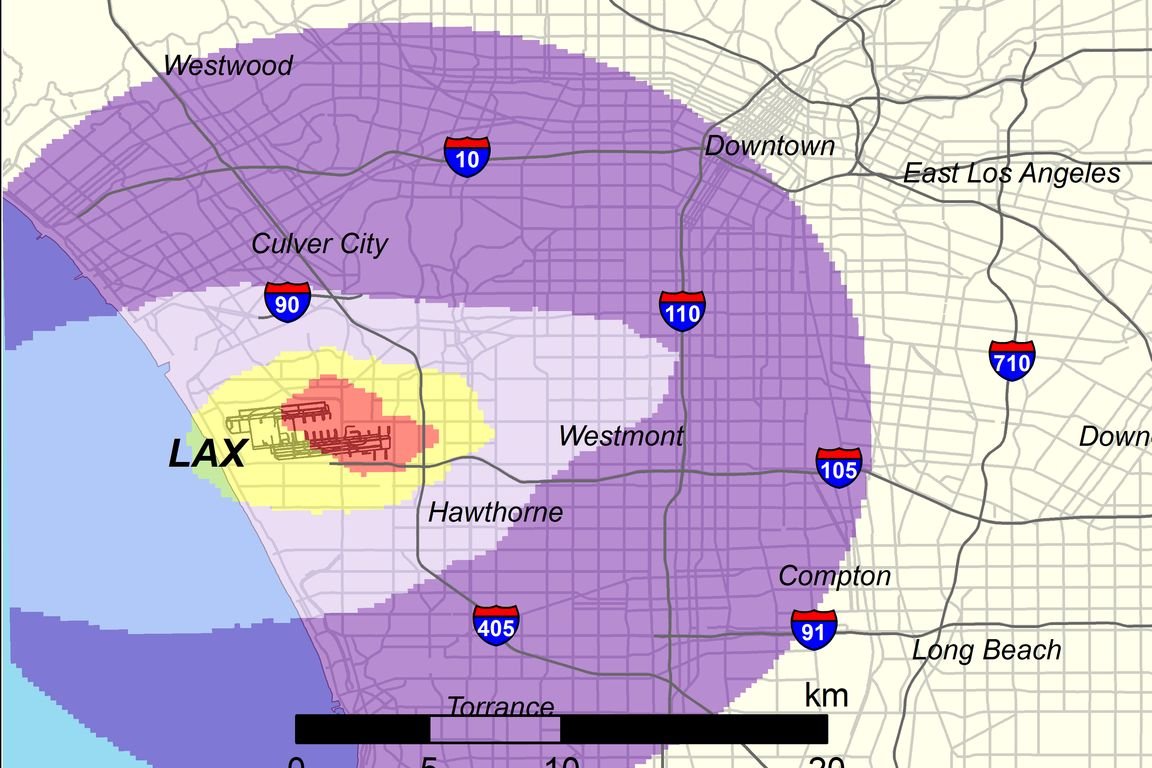
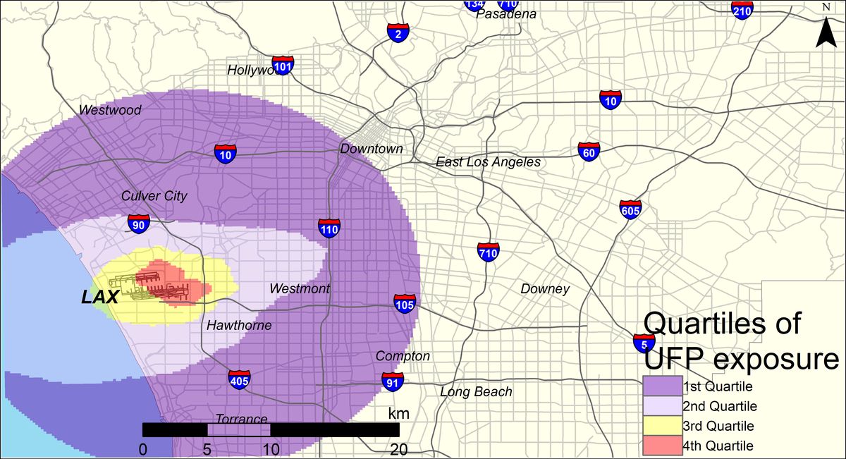
    UCLA researchers found that mothers who lived within the red area adjacent to LAX were 14% more likely to have a pregnancy that ended in a preterm birth. Those in the yellow area were 8% percent more likely than those in areas farther from the airport.

    A study from the UCLA Fielding School of Public Health has found that pregnant women exposed to high levels of ultrafine particles from jet airplane exhaust are 14% more likely to have a preterm birth than those exposed to lower levels.

    The researchers examined exposure among women living near Los Angeles International Airport, in an area that includes neighborhoods in Los Angeles, El Segundo, Hawthorne, Inglewood and several other communities inland from the airport.

    “The data suggest that airplane pollution contributes to preterm births above and beyond the main source of air pollution in this area, which is traffic,” said Dr. Beate Ritz, a professor in the departments of epidemiology and environmental health sciences at the Fielding School.

    Preterm birth is associated with complications such as immature lungs, difficulty regulating body temperature, poor feeding and slow weight gain.

    The research team, co-led by Ritz and Scott Fruin of the Keck School of Medicine at the University of Southern California, examined records for all births — a total of 174,186 —between 2008 and 2016 to mothers living within 9 miles (15 km) of LAX. They divided the overall area into four sections based on the amount of ultrafine particle, or UFP, pollution from jet exhaust, with the section nearest the airport experiencing the highest exposure.

    After adjusting for traffic-related air pollution and other variables that may affect the risk of preterm birth, including airport-related noise and the mother’s age, education level and race, they found that expectant mothers in the quarter with the highest average ultrafine particle exposure had 14% higher odds of a preterm birth than mothers in the quarter with the lowest exposure.

    “Nearly 2 million people live within a 10–mile radius of LAX, many of whom are exposed to elevated levels of aircraft-origin UFPs,” said Sam Wing, a scholar at the Fielding School who also worked on the study.

    The research is summarized in an article published July 22 in the journal Environmental Health Perspectives and is based on a lengthy study from the spring 2020 edition of the journal. Other authors include Timothy Larson and Sarunporn Boonyarattaphan of the University of Washington and Neelakshi Hudda of Tufts University.

    This research was supported by grants from the National Institute of Environmental Health Sciences, part of the National Institutes of Health.